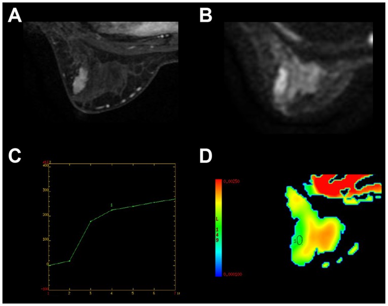
Figure 2.
Representative imaging results from a 35-year-old patient with fibroadenoma in the left breast. (A) The maximum diameter of the tumor was 2.6 cm, and demonstrated a well-defined, oval heterogeneously enhancing lesion on dynamic enhancement image. (B) The lesion was hyperintense on diffusion weighted imaging scans. (C) Time signal intensity analysis demonstrated a gradual progressive enhancement pattern (type 1 curve). (D) The lesion was hypointense on the ADC map with ADCmean=1.26×10⁻³ mm2/sec. ADC, apparent diffusion coefficient.

