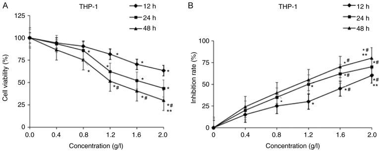
Figure 1.
Effects of matrine on the proliferation of acute myeloid leukemia cells. THP-1 cells were treated with 0, 0.4, 0.8, 1.2, 1.6 and 2.0 g/l matrine for 12, 24 and 48 h. Cell viability was assessed by a Cell Counting Kit-8 assay. (A) Cell viability rate. (B) Inhibition rate. Data are presented as the mean of at least three independent experiments. *P<0.05 vs. 0 g/l matrine; #P<0.05 vs. 0.4 g/l matrine; **P<0.05 vs. 1.2 g/l matrine.

