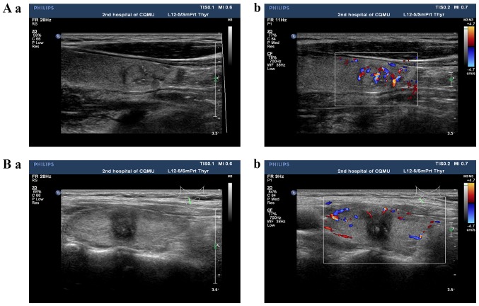
Figure 2.
Conventional ultrasound of papillary thyroid carcinoma using (Aa and Ba) 2-dimensional grayscale ultrasound and (Ab and Bb) a color Doppler ultrasound. (Aa and b) An isoechoic thyroid nodule with regular morphology, a clear boundary, an aspect ratio <1, the absence of microcalcification and a rich blood flow signal. (Ba and b) A hypoechoic thyroid nodule with irregular morphology, an unclear boundary, an aspect ratio ≥1, microcalcification and a poor blood flow signal. The red color in the center of the color Doppler ultrasound image indicates the blood flow signal of the nodule is toward the ultrasound probe, whereas the blue color indicates the opposite direction. The red and blue area on the right side of Ab and Bb indicates the blood flow range, which is the range of blood flow velocities that can be measured. Gray lines indicate the thyroid and the green lines indicate the position and direction of the ultrasound probe on the thyroid.

