Fig. 1.

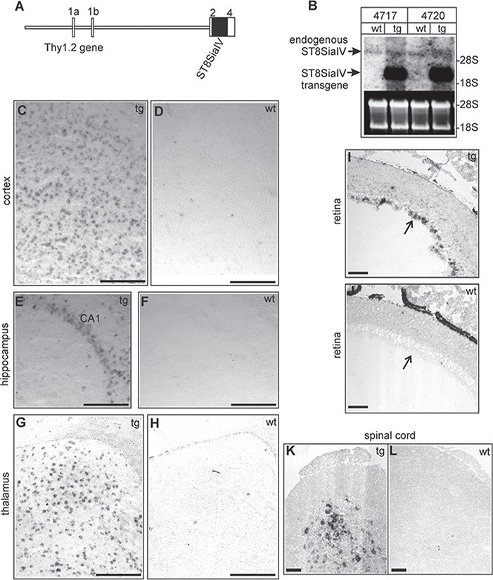
Generation of ST8SiaIV-tg mice. (A) Schematic representation of the tg construct. The murine ST8SiaIV cDNA was subcloned into the unique XhoI site of the Thy1.2 gene cassette (van der Putten et al., 2000) and is indicated by a solid box. Exons of the Thy1.2 gene are numbered 1a to 4 and are indicated by open boxes. (B) Northern blot analysis of tg mice of lines tg4717, tg4720 and wt littermates. Total RNA from tg and wt mouse brains (10 μg/lane) were separated by agarose gel electrophoresis, transferred to nylon membranes and hybridized to the ST8SiaIVspecific probe (entire coding sequence, black bar in A). Equal loading was controlled by ethidium bromide (EtBr) staining.(C–L) In situ hybridization of adult tg (linetg4717; tg) and wt was done on parasagittal brain sections, which were hybridized to ST8SiaIV specific antisense digoxigenin-labeled cRNA probes. Transgene expression was observed in the cortex (C), the hippocampal CA1region (E) and the laterodorsal thalamic nucleus (G). In wt mice only few cells gave hybridization signals (D, F, H). Hybridization of retina cryo-sections (age: P12) revealed transgene expression in the retina ganglion cell layer (arrow) in tg (I) but not in wt (J) mice. Transgene expression was also observed in the spinal cord of tg mice (K). ST8SiaIV expression was not detectable in wt spinal cord cross sections (L). (Control hybridizations with ST8SiaIV sense probes gave no specific hybridization signals (data not shown). Scale bars, 100 μm.
