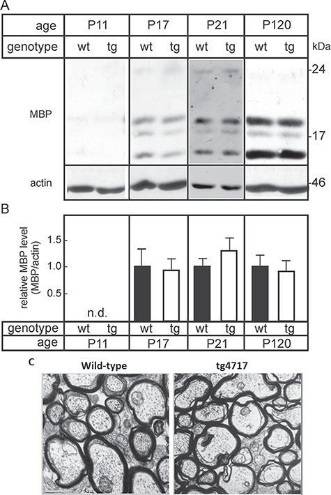
Fig. 3.

MBP expression in tg and wt mice. Forebrains were isolated from wt mice and tg littermates (line tg4717) at11, 17, 21 and 120 days of age. Total brain membrane fractions were prepared by centrifugation at 100,000 × g, separated (5 μg of protein/lane) in 12% SDS polyacrylamide gels and transferred onto nitrocellulose membranes. (A) Membranes were probed with antibodies against MBP and actin. (B) MBP bands were quantified by densitometry and normalized to actin protein levels. Mean MBP levels in wt brains were set to 100%. Columns indicate mean +/− SEM (n = 3 for P17and P120; n = 5 for P21) (n.d., not done). No significant differences between tg and wt mice were observed (t-test). (C) Electron micrographs of cross sections of the optic nerve did not show any obvious difference in the degree of myelination and myelin structure between tg mice (A) and wt littermates (B). Scale bar, 0.6 μm.
