Figure 6.
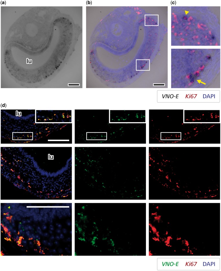
A lncRNA is expressed in vomeronasal progenitor cells. (a) Lower magnification microscopy black-and-white images of VNO sections stained by in situ hybridization for lncRNA VNO-E probe (dark staining). (b) Co-labelling for VNO-E probe (chromogenic detection) and Ki67 (fluorescent red staining). DAPI-stained nuclei are shown as blue overlaid fluorescence. (c) Higher magnification images of insets in centre panel showing striking localization near the base of the vomeronasal neuroepithelium (arrow) and around the corners (arrowhead). (d) Microscopy images of VNO sections subjected to double fluorescent in situ hybridizations for lncRNA VNO-E (green) and Ki67 (red). The first two rows of images depict co-localization between signals for the two genes in cells near the base of the epithelium, confirming the data presented in (a-c). The third row of pictures are high magnification images showing the co-localization of VNO-E and Ki67 in the vast majority of VNO-E-positive cells at the corners of the VNO neuroepitelium, where Ki67 staining is concentrated (neural progenitors). lu, VNO lumen. DAPI is pseudo-coloured in blue. Size bar is 100 μm.

