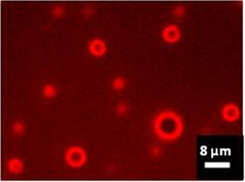
Figure 3.

Wide-field NIR fluorescence of ICG-labeled lipid MBs, 60 × oil immersion objective. The image was captured by a Nikon Inverted Microscope Eclipse Ti-E equipped with an LED source emitting at 770 nm (pE-100 LED, CoolLED Ltd., U.K.), a filter block containing an excitation filter, a dichroic beamsplitter (mirror) and a barrier/emission filter dedicated to ICG imaging (Chroma Technology, VT, U.S.A.), and a sCMOS camera (Zyla 4.2, Andor, U.K.).
