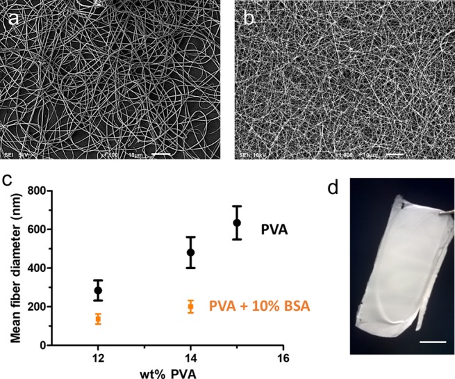
Figure 1.

Scanning electron microscope (SEM) images of electrospun samples: (a) 14 wt % PVA (scale bar 10 μm), (b) PVA/BSA (14 wt % PVA–10% BSA) (scale bar 10 μm), and (c) average fiber diameter (mean obtained over n = 100 measurements) measured for the PVA fiber (black dots) and the PVA loaded with 10 wt % BSA (orange squares). (d) PVA/BSA self-standing electrospun membrane (12 wt % PVA–10% BSA) as removed from the collector after electrospinning (scale bar 1 cm).
