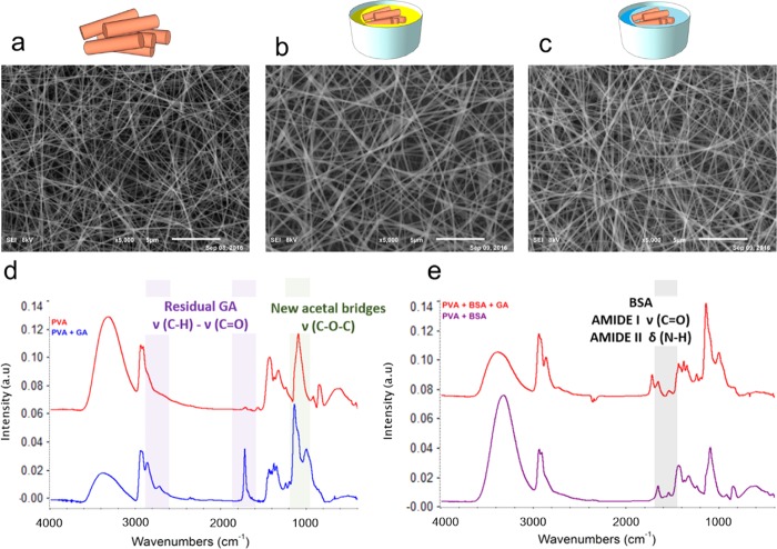
Figure 2.
Cross-linking of the PVA/BSA membrane with glutaraldehyde. (a) SEM micrograph of PVA 12 wt %–BSA 10% as-spun fibers (bar = 5 μm). (b) SEM micrograph of the sample after cross-linking with GA. The sample was immersed into an acetone bath containing 0.15 M glutaraldehyde and 0.05 M HCl for 1 h and gently dried (bar = 5 μm). (c) SEM micrograph of the cross-linked mat after overnight immersion in water and gentle drying (bar = 5 μm). (d) FTIR of a mat of PVA before (red line) and after (blue line) cross-linking with GA. (e) FTIR of a mat of PVA 12 wt %–BSA 10% before (purple line) and after (red line) cross-linking with GA. Relevant vibrational modes are highlighted for PVA and PVA/BSA, before and after cross-linking.

