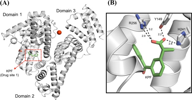
Figure 5.
Overall crystal structure of the BSA–ketoprofen complex and details of the KPF binding mode. (A) Cartoon representation of the BSA monomer showing the three-domain architecture of the protein and the drug site 1. Calcium ions and the ketoprofen molecule are represented by red spheres and green sticks, respectively. (B) Drug site 1 of BSA in complex with KPF. The hydrogen-bonding interactions between the ligand and the polar residues in the binding site are shown as dashed black lines. Distances are expressed in Ångstrom.

