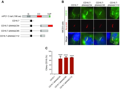
Figure 1.
A 42-residue fragment at the C terminus of PC1 likely harbors a new CTS. A) Schematic representation of CD16.7 with and without different PC1 C-terminal fragment fusions. B) Expression of indicated constructs in A in the primary cilia of IMCD3 cells. Cells were stained by antibodies against CD16 (green) and acetylated α-tubulin (Ac-α-tub; red). Scale bar, 5 μm. C) Quantification of percentage of CD16-positive cilia in cells. A total of ≥50 ciliated, transfected cells were counted for the presence of CD16 signal on cilia under each condition. Error bars represent the sd between microscope fields from ≥3 independent experiments. G, G-protein activation domain; CC, coiled-coil domain. ****P < 0.0001 compared with CD16.7 control.

