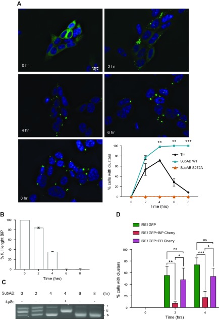
Figure 3.
Formation of large clusters after BiP cleavage. A) BiP cleavage induces massive and irreversible clustering. HAP1KO cells expressing WT IRE1GFP were treated with SubAB WT or S272A mutant (0.1 μg/ml) for the indicated times. Representative fields recorded at the indicated times are shown together with a quantitation of the percentage of cells with clusters over the time course. At least 100 cells were evaluated per time point and statistically significant differences between Tm and SubAB WT time points are indicated. **P < 0.01, ***P < 0.001. Scale bar, 10 μm. B) Time course of SubAB cleavage of BiP. Cells expressing IRE1GFP WT were treated with SubAB (0.1 μg/ml) for the indicated times. Proteins were then extracted and analyzed by Western blot with anti-BiP antibody. One of the blots is shown in Supplemental Fig. S4A. 14.3.3 was used as housekeeping. The full-length (∼78 kDa) and cleaved BiP (∼28 kDa) bands were quantified using ImageJ and the percentage of full-length BiP is plotted. C) SubAB induces XBP1 splicing. HAP1KO cells expressing WT IRE1GFP were treated with SubAB (0.1 μg/ml) for the indicated times. XBP1 mRNA splicing was determined by RT-PCR, % XBP1 splicing was calculated and plotted. Note that the activation of IRE1α persists throughout the experiment and is inhibited by 16 μM 4μ8c treatment, demonstrating IRE1α dependence. D) Overexpression of BiP inhibits Tm-inducible clustering of WT IRE1GFP. HEK 293T cells expressing Tet-inducible IRE1GFP WT were transiently transfected with either BiP-mCherry or ER-mCherry and treated with Tm (4 μg/ml) for the indicated times. Cells with clusters that were positive for mCherry were counted and graphed. At least 88 cells were evaluated, and experiments were performed at least 3 times. Ns, not significant; P > 0.05, *P < 0.05, **P < 0.01, ***P < 0.001.

