Figure 1.
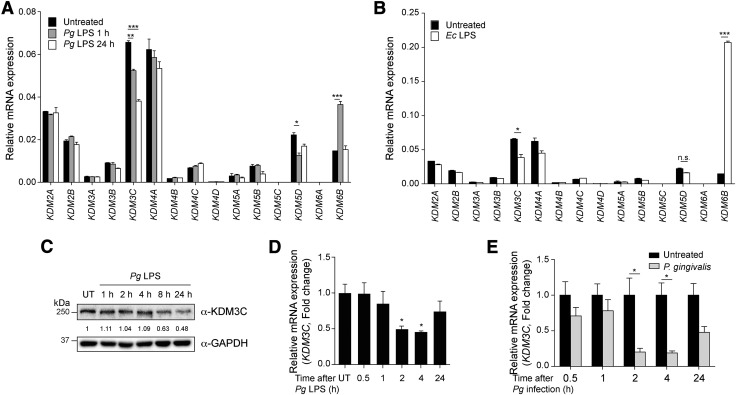
KDM3C is negatively regulated by Pg and its LPS treatment in THP-1 cells. A, B) The mRNA expression profiling of KDMs in PMA-differentiated THP-1 cells treated with Pg LPS (1 µg/ml, 1 and 24 h) (A) or Ec LPS (1 µg/ml, 1 h) (B). The mRNA expression of KDMs was assessed by using real-time qPCR. Expressions values were normalized by GAPDH. C) Changes in the levels of KDM3C protein expression was determined by Western blotting with time-dependent Pg LPS (1 µg/ml) exposure in THP-1 cells. Molecular mass markers are indicated. GAPDH was used as a loading control. KDM3C and GAPDH fold change values, which were normalized to untreated (UT) control, were obtained by using densitometry. D) KDM3C mRNA expression in Pg LPS–treated THP-1 cells was determined by real-time qPCR. E) THP-1 cells were infected with Pg for the indicated time points, and the expression of KDM3C mRNA was determined by real-time qPCR. Data are representative of at least 2 or more independent experiments and were statistically analyzed by Student’s t test; n.s., not significant. *P < 0.05, **P < 0.01, ***P < 0.001 compared with the untreated controls.

