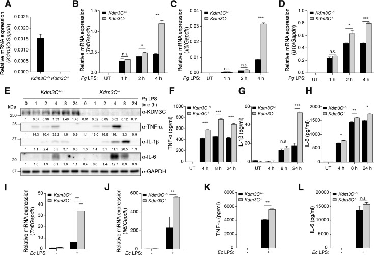
Figure 3.
Deletion of KDM3C shows the increased inflammatory response to Pg LPS in BMDMs. A) BMDMs from Kdm3C WT (Kdm3C+/+) and KO (Kdm3C−/−) mice were cultured, and real-time qPCR was used to measure endogenous Kdm3C mRNA levels. B-D) Real-time qPCR analysis of mRNA from Kdm3C+/+ and Kdm3C−/− BMDMs treated with Pg LPS (1 µg/ml) as indicated. mRNA expression levels of cytokines, Tnf (B), Il6 (C), and Il1b (D), were determined. E) Western blotting analysis of the levels of indicated proteins in Kdm3C+/+ and Kdm3C−/− BMDMs treated with Pg LPS at indicated times. GAPDH was used as a loading control. Protein expression was quantified, and the ratio of normalized protein is shown as fold change. F–H) Secretions of cytokines TNF-α (F), IL-1β (G), and IL-6 (H) into the culture supernatant from Kdm3C+/+ and Kdm3C−/− BMDMs after stimulation of Pg LPS (4, 8, 24 h) were measured by ELISA. I–L) mRNA expression levels of Tnf (I) and Il6 (J) and secretion levels of TNF-α (K) and IL-6 (L) were determined upon Ec LPS (1 µg/ml) exposure in Kdm3C+/+ and Kdm3C−/− BMDMs. UT, untreated. Data are representative of at least 3 independent experiment and statistically analyzed by Student’s t test; n.s., not significant. *P < 0.05, **P < 0.01, ***P < 0.001 compared with WT cells.

