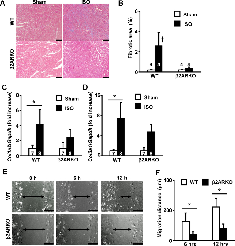
Fig 2. Deletion of β2AR attenuates ISO-induced cardiac fibrosis and migration of cardiac fibroblasts.
(A) Representative images of M-T staining of heart tissue obtained from wildtype or β2ARKO mice at 2 weeks after sham-operation or ISO-stimulation. The bar indicates 100 μm. (B) Quantification of fibrotic area assessed by M-T staining. (C, D) Expression levels of cardiac fibrotic marker gene, Col1a2 (C) and Col3a1 (D) in ventricles determined by real-time RT-PCR. Values were normalized to that of GAPDH and are represented as fold increases relative to that in the wildtype sham group. (E) Representative images of cell migration as assessed by scratch assay. Images of scratched confluent cardiac fibroblasts from wildtype control (upper panels) or β2ARKO (lower panels) mice were taken at indicated time points. Arrows indicate the width of cell-free areas. The bar indicates 200 μm. (F) Quantitation of migration distance of cardiac fibroblasts at 6 h and 12 h. Values are mean ± SD, and the number displayed on each column indicates the number of samples. †P<0.05 vs. all other groups, *P<0.05 between two indicated groups by one-way ANOVA followed by Tukey-Kramer test.

