
Fig. 5. Loss of LSEC autophagy amplifies liver fibrosis without increasing liver injury.
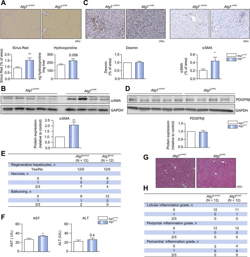
Atg7endo and Atg7control mice were treated every other day with CC14 i. p. for 1 week to induce mild acute liver injury (n = 24). (A) Whole liver sections stained for Sirius Red and quantification of Sirius Red-positive area (left) and Hydroxyproline stain content (right). (B) Immunoblots for αSMA in isolated HSCs from Atg7endo and Atg7control mice and protein quantification. (C) Whole liver sections stained for desmin and αSMA and quantification of positive area. (D) Immunoblots for PDGFRB in whole liver from Atg7endo and Atg7control mice and protein quantification. (E) Histological liver analysis for hepatocyte regenerative capacity and liver injury. (F) Aminotransferase levels showing an increase in aspartate aminotransferase with no change in alanine aminotransferase. (G) Representative images of whole liver sections stained for H&E and (H) histological liver analysis for inflammation scoring in Atg7endo and Atg7control mice after mild acute liver injury (CCl4 i.p. for 1 week). Representative images are shown. Data shows mean value ± SEM of at least 3 experiments (*p ≤0.05, **p ≤0.01, ***p ≤0.001, Student’s t test). CCl4, carbon tetrachloride; H&E, hematoxylin and eosin; HSC, hepatic stellate cell; LSEC, liver sinusoid endothelial cell.