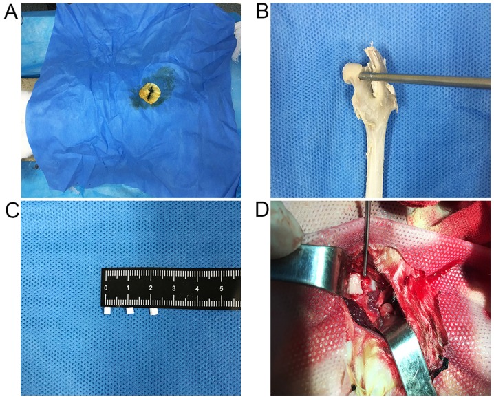
Figure 2.
CD and graft filling. (A) After successful anesthesia, the incision was marked, the operative area was disinfected and the sterile opening was covered with a towel. (B) Demonstration of the CD approach to the femoral head in vitro. (C) nHAC/PLA scaffold size measurement (2 mm3). (D) nHAC/PLA or BMSCs-nHAC/PLA were implanted into the decompression tunnel by fit pressure. CD, core decompression; BMSC, bone marrow stem cells; nHAC/PLA, nano-hydroxyapatite/collagen I/poly-L-lactic acid; H&E, hematoxylin and eosin; CT, computed tomography; ANFH, avascular necrosis of the femoral head; MRI, magnetic resonance imaging.

