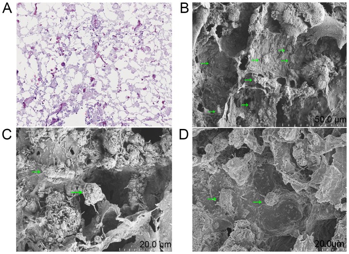
Figure 3.
BMSCs could attach to scaffolds. (A) A total of 24 h after seeding, hematoxylin and eosin staining showed that a large number of BMSCs adhered to the scaffold and were distributed evenly (×200). (B) Scanning electron microscopy micrographs revealed that BMSCs were uniformly distributed inside the scaffold; the cell bodies and filamentous pseudopodia contacted the micropore wall. (C) The bodies of the poorly attached cells were spherical in shape and only two to three short and thick pseudopods contacted the inner surface of the scaffold. (D) The adherent cell bodies were long and oval-shaped, or spherical. One side of which was closely attached to the pore wall of the scaffold and many banded pseudopodia radiated from it (denoted by the green arrowhead). BMSCs, bone marrow stem cells.

