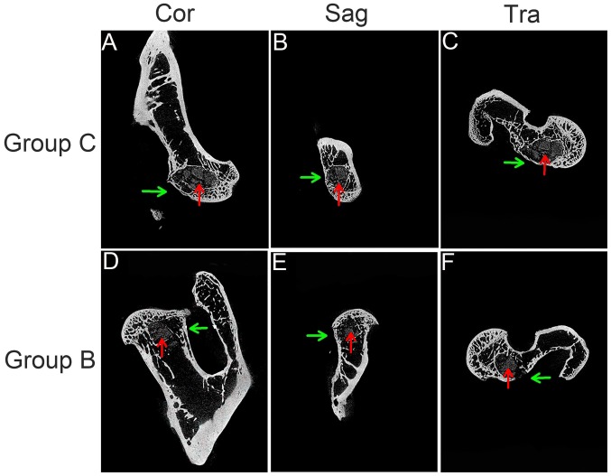
Figure 5.
Micro-CT examination. (A) Cor, (B) Sag and (C) Tra micro-CT examination showed that the trabeculae around the subfemoral decompression tunnel in group C were compact and arranged regularly, the new bone gradually grew into the center of the tunnel, and tissue-engineered bone was wrapped well and degraded. At the same time, the exit of the tunnel was sealed by a thin layer of new bone. (D) Cor, (E) Sag and (F) Tra micro-CT examination showed that the trabeculae around the subfemoral decompression tunnel in group B were loose and disordered. No new bone was found in the inner wall and scaffold of the tunnel, and the degraded nHAC/PLA was not tightly wrapped by the new bone, and the exit of the tunnel was not sealed by the new bone (green arrows point to decompression tunnels and red arrows point to implants). CT, computerized tomography; Cor, coronal section; Sag, sagittal section; Tra, transverse section; nHAC/PLA, nano-hydroxyapatite/collagen I/poly-L-lactic acid.

