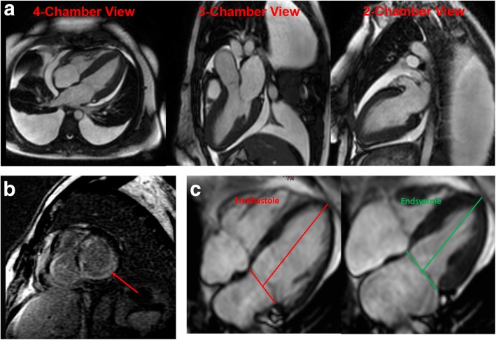
Fig. 1.
Representative cardiovascular magnetic resonance (CMR) images of a) a patient with light chain (AL) amyloidosis demonstrating global left ventricular (LV) wall hypertrophy, pericardial effusion and both-sided pleural effusions, b Late gadolinium enhancement (LGE) pronounced in the subendocardial layers in cardiac amyloidosis (marked with a red line) and c) long axis strain (LAS) measurement

