Fig. 1.
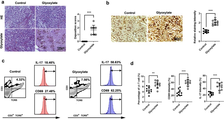
CaOx crystals activate γδT cells in the kidney. Ten mice were intraperitoneally injected with glyoxylate at 100 mg/kg or 0.9% saline once daily for 7 days. a HE and von Kossa staining was performed in mouse kidneys. Deposition scores were measured. b Immunohistochemical staining of TCRγδ in kidneys. The staining intensity of TCRγδ was calculated in 10 high power microscopic fields. c γδ T (CD3+, TCRγδ+) cells were gated and tested for the expression of CD69/IL-17 by flow cytometry. d The percentage of γδT cells and the expression of CD69 and IL-17 gated from γδT cells were quantified by flow cytometry (10 mice per group). *P < 0.05; **P < 0.01; ***P < 0.001

