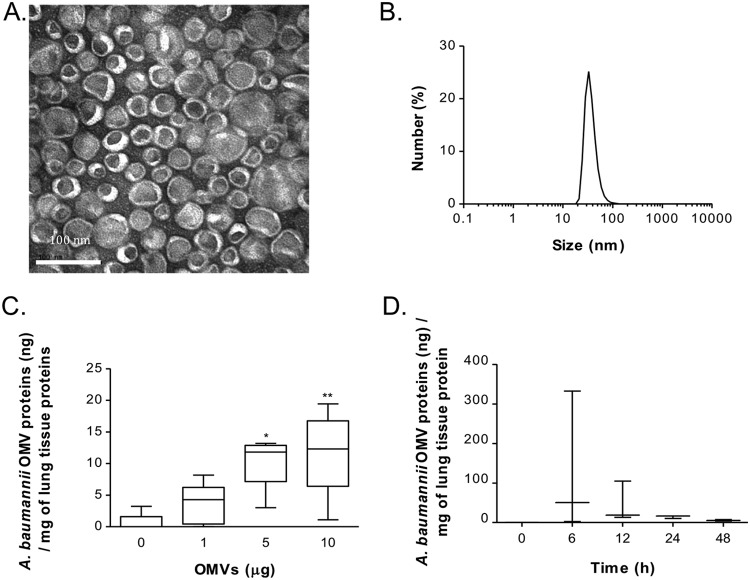
FIG 1.
Characterization of A. baumannii OMVs and OMV component detection in the lungs. (A) Transmission electron microscopy indicating that the purified OMVs have lipid bilayered structures. (B) Size distribution of the purified OMVs determined by dynamic light scattering, with the diameter ranging from 30 to 100 nm. (C) Various amounts of A. baumannii OMVs (0, 1, 5, and 10 μg in total protein amounts) were intranasally introduced into mice, and the amounts of OMV components were measured at 24 h after intranasal instillation of the OMVs. (D) A. baumannii OMVs (10 μg) were intranasally introduced to mice, and the amounts of OMV components were measured at 0, 6, 12, 24, and 48 h after intranasal instillation of the OMVs. *, P < 0.05; **, P < 0.01.

