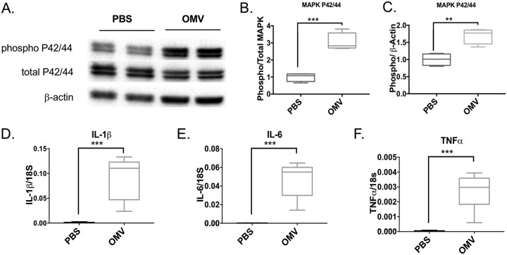
FIG 7.
OMVs activate MAPK and NF-κB pathways in the mouse lung. C57BL/6 mice were intranasally administered A. baumannii OMVs (5 μg in total protein amounts) or PBS as a control. Mice were sacrificed at 6 h postintranasal administration of OMVs to harvest lung samples. MAPK activation was measured by Western blot analysis of lung samples. (A) A representative Western blot image. (B and C) Densitometry analysis of band intensity was performed. The levels of phospho-MAPK were normalized to total MAPK (B) or β-actin (C). The NF-κB activation was measured by activation of downstream genes in the NF-κB pathway. (D to F) The expression levels of IL-1β (D), IL-6 (E), and TNF-α (F) were measured by qPCR. P < 0.01 (**) and P < 0.005 (***) compared to the control group. n = 6 mice in each group.

