Fig. 2.
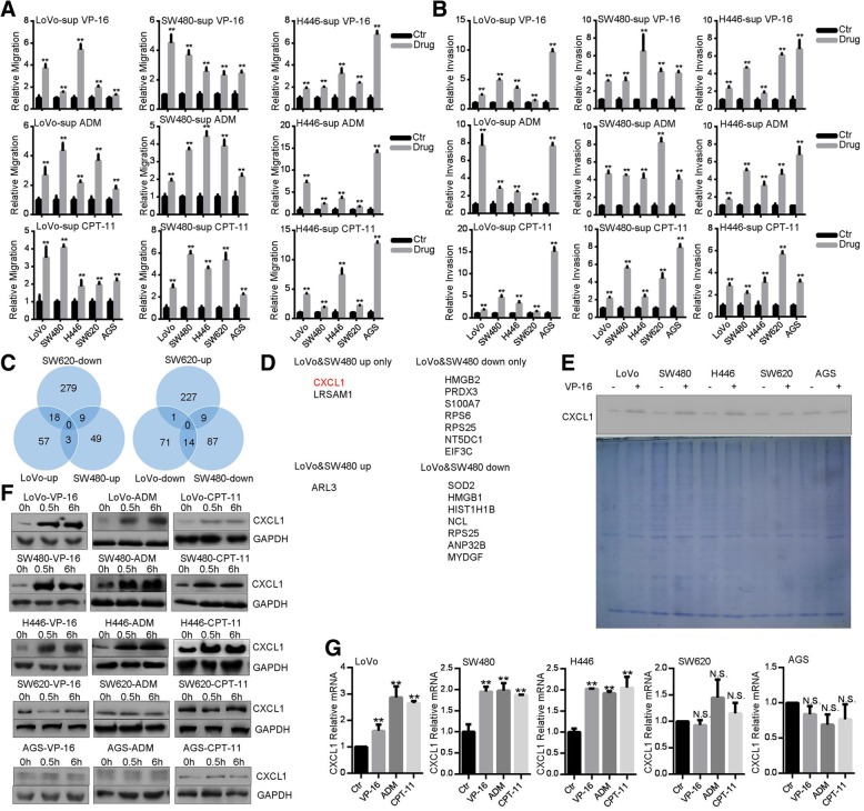
Topoisomerase inhibitors promote CXCL1 expression and secretion. a, b CM from TI-treated cells promotes migration (a) and invasion (b). Cells (LoVo, SW480, H446) were treated with VP-16 (20 μM), CPT-11 (80 μg/ml), or ADM (0.2 μg/ml) for 0.5 h in complete medium. After removing medium and washing with PBS, cells were cultured in serum-free culture medium for 4 h. CM was collected and used for transwell assays for indicated cells. c Wynn diagram of differential protein expression profiles in CM of LoVo, SW480 and SW620 cells. d The list of differentially expressed proteins. e Western blot analysis of CXCL1 in the CM. Indicated cells were treated with VP-16 (20 μM) for 0.5 h in complete medium, and CM was collected as in (a, b). Part of CM was resolved by SDS-PAGE and gel was stained with coomassie blue. f Western blot analysis of CXCL1 expression in cells after treatment with VP-16 (20 μM), CPT-11 (80 μg/ml), or ADM (0.2 μg/ml) for 0.5 and 6 h. g qRT-PCR analysis of CXCL1 mRNA after treatment with VP-16 (20 μM), CPT-11 (80 μg/ml), or ADM (0.2 μg/ml) for 0.5 h

