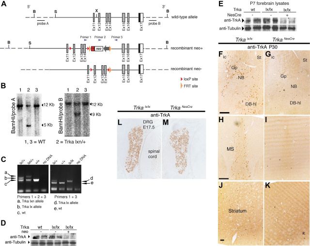
Figure 2.
Generation of CNS-specific Trka conditional mice. A, Trka conditional targeting strategy. Schematic representation of exons 11–17 of the wild-type mouse Trka locus; recombinant (neo+) and recombinant (neo−) alleles are also shown. LoxP sites flanked the Trka exons 12–14, whereas FRT sites flanked the neo gene, which was subsequently excised in vivo by crossing the Trka floxed mice with transgenic mice expressing Flp recombinase ubiquitously (Rodríguez et al., 2000). This resulted in the generation of the recombinant Trkalx_neo− (Trkalx) allele. B, Southern blot analysis of targeted ES cell clones of Trkalx_neo+(Trkalxn) using two probes at the 5′ and 3′ sides of the targeting construct, as indicated. BamHI restriction enzyme was used to digest the genomic DNA for the Southern blot analyses. C, PCR analysis of Trkalxn and Trkalx alleles. The positions of the primers used for PCR analysis are indicated in A. The PCR reaction with the primers 1 and 3, which showed successful deletion of the neo gene, was used for routine genotyping of the Trkalx mice. D, Western blot analysis of gp140TrkA protein in forebrains of newborn (P1) wild-type, Trkalx/lx, and Trkalxn/lxn mice. While TrkA protein level was nearly not detectable in forebrain lysates of Trkalxn/lxn mice, removal of the neo cassette restored normal TrkA protein level in Trkalx/lx mice compared to control mice. Tubulin was used to control for loading. E, Western blot analysis showing Cre-mediated removal of TrkA from P1 forebrain lysates of Trkalx mice crossed to NestinCre mice. While TrkA protein levels in wild-type and Trkalx/lx mice were identical, no expression of TrkA was detected in the Trkalx/lx;NesCretg/+ mice (TrkaNesCre). F–K, Forebrain coronal cryosections from 1-month-old TrkaNesCre and control (Trkalx/lx) mice were stained with antibody against TrkA. Although strongly stained neurons were found in all the indicated areas in the control mice, shown is mainly the nucleus basalis (F, G), the medial septum (H, I), and the striatum (J, K); no TrkA-positive cells were found in the TrkaNesCre mice. L–M, Transversal cryosections of the spinal chord region of E17.5 mouse stained with TrkA antibodies. Clear TrkA-positive staining is observed in neurons of the DRG in both control and TrkaNesCre mice. St, Striatum; ic, internal capsule; Gp, globus pallidus; DB-hl, diagonal band horizontal limb. Scale bars: F (for F, G), H (for H, I), 200 μm; J (for (J, K), 100 μm. B, bamHI; S, SacII; X, XhoI.

