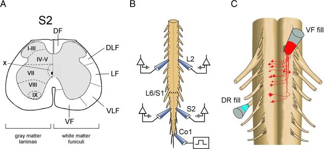
Figure 1.
Studies of sacral neurons with rostral projections through the VF. A, Schematic cross section through the S2 segment of the spinal cord. VF, VLF, LF, DLF, and DF are ventral, ventrolateral, lateral, dorsolateral, and dorsal funiculi, respectively. B, Illustration of the isolated en bloc spinal cord preparation is shown with the recording electrodes from the left and right S2 and L2 ventral roots. The first coccygeal dorsal root (Co1) is stimulated to produce the rhythm. A hyphenated line indicates the lumbosacral junction (L6–S1). C, Primary afferent innervation of VF neurons. Retrograde labeling of VF neurons through cut VF axon bundles at the lumbosacral or the S1–S2 junction (red) and anterograde labeling of contralateral afferents entering the sacral cord through the S3 dorsal root (cyan) by different fluorophores. The en bloc spinal cord preparation is shown with ventral side up. Vibratome cross sections of the fixed preparation were immunostained for VGluT1 or VGluT2 to determine a possible glutamatergic innervation of VF neurons by SCA, as described in Figures 2, 3, and 4.

