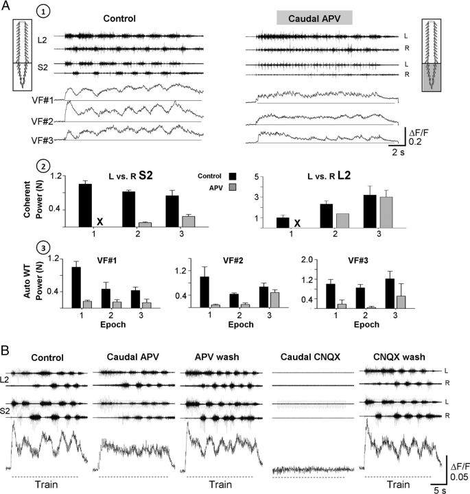
Figure 10.
The involvement of rhythmic and nonrhythmic VF neurons and the sacral CPGs in sensory activation of the hindlimb CPGs. A, The activity of VF neurons and the motor output recorded from the left and right ventral roots of S2 and L2 during 40-pulse 5-Hz stimulus trains applied to the Co1 dorsal roots at 12 μA, are shown before (1; Control) and 15 min after (1; Caudal APV) application of the NMDA receptor antagonist APV (20 μm) to the sacral segments in a dual-chamber experimental bath. The top 2 histograms (A2) show the normalized power of the motor rhythm (mean ± SD), produced by SCA stimulation and recorded from the left and right ventral roots of S2 and L2 before (A2; Control, black bars) and after addition of APV to the sacral compartment (A2; APV, gray bars). The mean power for each pair of time series (L vs R S2, and L vs R L2) was calculated at the beginning, middle, and end of the train (epochs 1, 2, and 3, respectively). A3, The mean ± SD of the normalized auto-power of the ΔF/F of the 3 imaged neurons (VF 1, 2, and 3) under these conditions. The normalized cross-power and auto-power values were calculated from the high-power frequency bands of the respective cross-power and auto-power density plots obtained by WT analyses after coherence test and 95% significance test of a χ2 power distribution, respectively (for further details, see Results). B, The rhythmic activity from an RM-type VF neuron and the motor output recorded from the left and right S2 and L2 ventral roots during stimulation of the Co1 dorsal root are shown before (Control), 20 min after addition of APV to the sacral segments (Caudal APV), 35 min after APV wash (APV wash), 10 min after addition of 10 μm CNQX to the sacral segments (Caudal CNQX), and 60 min after CNQX wash (CNQX wash). Hyphenated lines indicate trains. Note that the rhythmic response of the VF cells is attenuated by APV, recovers when APV is washed, and all the activities are abolished by CNQX. Stimulation parameters: 50-pulse 2.5-Hz trains were applied at 10 μA. A, B, The neurons studied are right-S2 neurons back-labeled from the contralateral VF at the lumbosacral junction.

