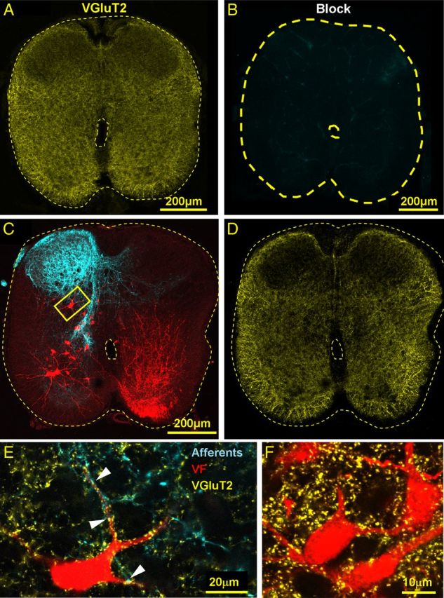
Figure 3.

Primary afferent and intraspinal VGluT2 IR innervation of VF neurons. A, B, The confocal micrograph in A visualizes the specific immunostaining for VGluT2 (yellow) in a cross section through the S2 segment, which is completely abolished by pretreatment with the blocking peptide of the primary VGluT2 antibodies (B). C, Low-power projected confocal micrographs of a 50-μm cross section cut through the S2 segment of the spinal cord shows VF neurons retrogradely labeled through the left VF at the S1–S2 level (red) and anterogradely filled sacral afferents (cyan) labeled via the contralateral (right) S3 dorsal root. D, The low-power cross section displayed in C is shown after VGluT2 immunostaining (yellow). E, High-power (original magnification × 60), single-slice micrograph of the area framed in C shows a VF neuron (pseudo color, red), which is contacted (e.g., arrowheads) by primary afferent boutons (pseudo color, cyan) with VGluT2 IR (yellow). Fluorophores: VF, Cascade blue dextran; Afferents, Texas red dextran; and VGluT2, Cy5. F, High-power (original magnification × 60) view of a single optical slice micrograph of a group of S2 VF neurons back-labeled in a different double labeling experiment followed by immunostaining for VGluT2. The neurons are innervated by VGluT2 IR boutons but are not contacted by SCA.
