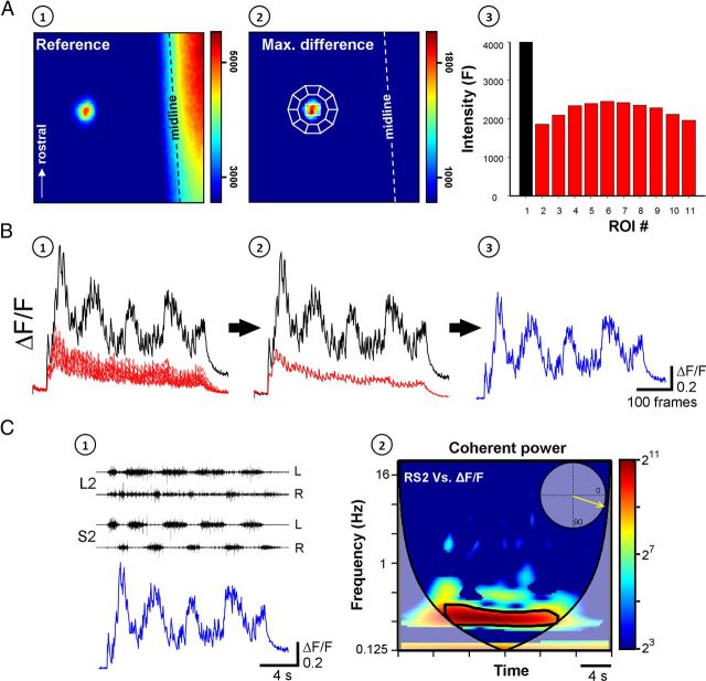
Figure 6.
The activity of VF neurons and its correlation to the concurrent motor output are used to evaluate the involvement VF neurons in sensory activation of the CPGs. A, A single VF neuron, in the right S2 segment, back-labeled with Calcium green dextran from cut VF axon bundles at the contralateral (left) lumbosacral junction and imaged from the ventral surface of the en bloc spinal cord, is shown before (A1, reference image) and during SCA stimulation (A2, maximal difference image). The neuron is outlined by a white line and encircled by 10 regions of interest used to measure the out-of-focus fluorescence. The graph (A3) presents the mean pixel intensity of the neuron (black) and the encircling ROIs (red) calculated from a five frame average taken before the stimulus train. Arrow indicates the rostral direction. B, The relative fluorescence changes (ΔF/F) of the neuron (black) and the encircling ROIs (red) during the stimulus train are shown in B1. The specific ΔF/F of the neuron (blue, B3) was calculated by subtracting the mean ΔF/F of the encircling ROIs (red, B2) from that of the neuron (black, B2). C, The rhythm produced by stimulation of the Co1 dorsal root and recorded from the left and right ventral roots of S2 and L2 is superimposed with the interpolated ΔF/F of the imaged neuron, as shown in C1. The coherent cross-power density plot of the activity recorded from the right S2 ventral root versus the ΔF/F of the imaged neuron is shown in C2. The mean R-vector in the circular inset plot, which was calculated from the high-power frequency band delineated by the black line, shows that both rhythms (right VF neuron and right S2 ventral root) are in phase. Stimulation parameters: 40-pulse 3-Hz train applied at 9 μA. Imaging was at 27 fps, the fluorescence excitation at 488 nm, and emission measured at 530 ± 15 nm. This figure describes the strategy of the series of experiments combining fluorescence and electrical activity measurements.

