Figure 1.
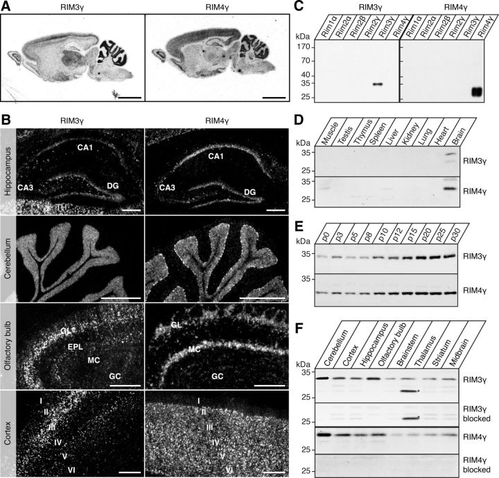
Overlapping but distinct expression patterns of RIM3γ and RIM4γ in adult rat brain. A, ISH micrographs showing RIM3γ and RIM4γ mRNA distribution in the whole brain. Negative controls with excess unlabeled oligonucleotides were devoid of signal (results not shown). Scale bar, 5 mm. B, Higher resolution pictures of emulsion-dipped sections of the hippocampus, the cerebellum, the olfactory bulb, and the cortex for RIM3γ (left) and RIM4γ (right). Scale bar, 300 μm. C, Homogenates of HEK-293T cells transfected with the indicated full-length RIM expression plasmids were analyzed by immunoblotting with affinity-purified antisera against RIM3γ and RIM4γ. The two antibodies were specific for the respective isoforms (peptides) they were raised against. D, Western blot analysis of adult rat tissues. Specific bands corresponding to the molecular weight of RIM3γ (32 kDa) and RIM4γ (27 kDa) were only detected in brain. E, Whole-brain homogenates from rats of the indicated ages (P0–P30) analyzed by immunoblotting with specific antibodies against RIM3γ and RIM4γ. Expression of both isoforms increased during postnatal brain development. F, Homogenates from the specified brain regions were analyzed by immunoblotting with isoform-specific antibodies and specificity of the antibody controlled by peptide blocking. Incubation of the blot with RIM3γ antibody revealed an unspecific 26 kDa band (*) in the thalamus. RIM3γ and RIM4γ proteins were ubiquitously expressed in the brain. However, the expression levels differed between the isoforms in various brain regions. GL, glomerular layer; EPL, external plexiform layer; MC, mossy cell; GC, granular cell; DG, dentate gyrus; I–VI, cortical layers.

