Figure 10.
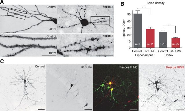
RIM3γ in vivo rescue of neuronal morphology, reduction in spine density after loss of RIM3γ in hippocampus and cortex. A, Hippocampal neurons in P21 rats show that neurons expressing the shRNA against RIM3γ exhibit a reduced number of spines compared with control cells expressing only RFP. B, Quantification revealed a significant loss in spine density after knock-down of RIM3γ in hippocampal and cortical neurons (t test, hippocampus ***p = 0.0003, cortex **p = 0.0042). C, Lentiviral particles expressing RFP (Control) and the shRNA against RIM3γ alone (shRIM3) or together with a green fluorescent-resistant variant of RIM3 (Rescue RIM3) were injected into P0 rat brains. At P21, RIM3 knock-down cortical neurons exhibited the expected loss in arborization, while neurons expressing both shRNA and the resistant RIM3 were undistinguishable from control neurons expressing RFP. Scale bar, 50 μm.

