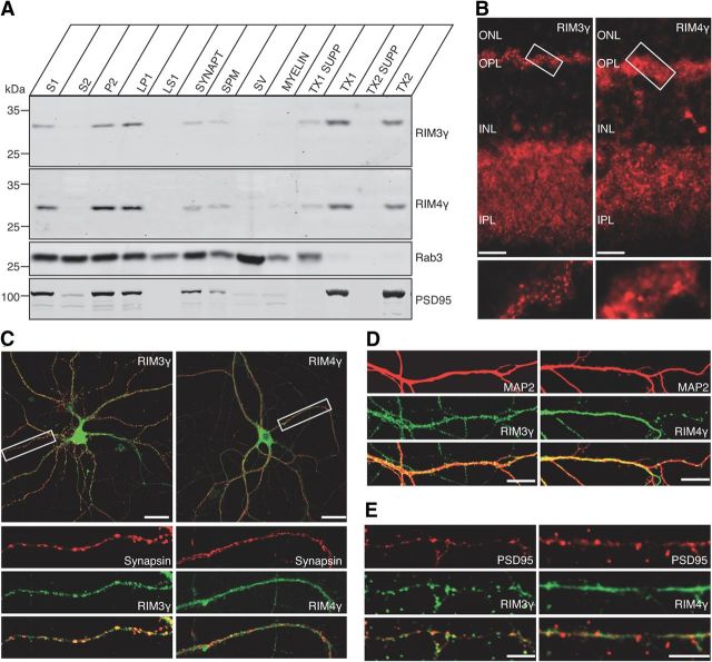
Figure 2.
RIM3γ and RIM4γ proteins are components of the presynaptic and postsynaptic cytomatrix. A, Rat brain homogenates were fractionated into the crude synaptosomal fraction (S1), the synaptosomal cytosol fraction (S2), the crude synaptosomal pellet fraction (P2), and the lysed synaptosomal membrane fraction (LP1, LS1), which consists of synaptosomal cytosol and SV-enriched fraction: the crude SV, the SPM, and myelin. The SPM was extracted twice with increasing Triton X-100 concentrations yielding the supernatant of the 0.5% (w/v) or 1% (w/v) Triton X-100 soluble fraction (TX1 SUPP and TX2 SUPP, respectively) and the Triton X-100 insoluble fraction of the SPM (TX1 and TX2). Fractions were analyzed using antibodies against RIM3γ and RIM4γ, as well as against Rab3A and PSD-95. Even though a fraction of RIM3γ and RIM4γ is extracted by Triton X-100, a substantial amount of the two proteins is still associated with the Triton X-100 insoluble fraction after the second extraction, resembling the pattern observed with PSD-95. B, Confocal micrographs of vertical rat retina sections labeled with antibodies against RIM3γ or RIM4γ. ONL, outer nuclear layer; OPL, outer plexiform layer; INL, inner nuclear layer; IPL, inner plexiform layer. Scale bar, 10 μm. RIM3γ exhibits a specific labeling in both synaptic layers, whereas RIM4γ is also present at these synapses but more broadly distributed. C–E, Double immunolabelings of hippocampal neurons (DIV 14) with the presynaptic marker Synapsin (C), the dendritic marker MAP2 (D), and the postsynaptic marker PSD-95 (E). Scale bars: (in C) 30 μm; (in D, E)10 μm.

