Figure 3.

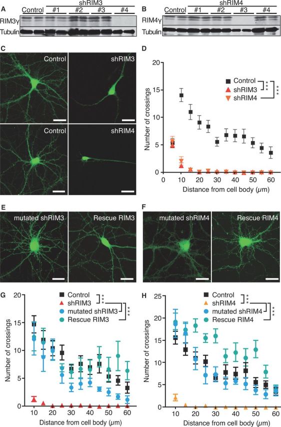
Knock-down of RIM3γ and RIM4γ results in altered neuronal morphology. A, B, Immunoblotting of cellular lysates from hippocampal primary neurons (DIV 14) transduced on DIV 1 with lentiviral particles expressing various RIM3γ-specific (A) or RIM4γ-specific (B) shRNAs (SH#1–4) and the empty vector (Control) revealed that shRIM3 results in a strong reduction of RIM3γ, and shRIM4 strongly reduces RIM4γ protein levels. Staining against tubulin was used as loading control. C, Hippocampal neurons were transduced at DIV 1 with lentiviral particles expressing GFP and either the empty vector (Control) or the RIM3γ/RIM4γ shRNA (shRIM3/4). All neurons were analyzed at DIV 14 using confocal microscopy. D, Sholl analysis revealed a loss of neuronal processes. E, F, The specificity of the observed phenotype was examined by transfecting either mutated shRNAs (mutated shRIM3/4) or functional shRNAs together with resistant RIM3γ/RIM4γ containing silent mutations (Rescue RIM3/Rescue RIM4). G, H, Quantification of the experiments in E and F by Sholl analysis for RIM3γ (G) and RIM4γ (H). Together, these control experiments show that the observed phenotype caused a reduction in the levels of RIM3γ and RIM4γ. One-way ANOVA, ***p < 0.001.
