Figure 4.

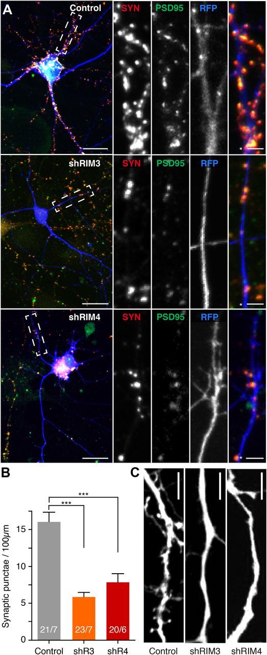
Absence of dendritic spines and reduction in synapse density in RIM3γ and RIM4γ knock-down neurons. A, Hippocampal neurons transfected at DIV 3 with either a vector-expressing GFP (Control) or GFP and the shRNA against RIM3γ (shRIM3) or RIM4γ (shRIM4). All neurons were immunostained using anti-Synapsin (SYN) and anti-PSD-95 (PSD-95) antibodies and analyzed at DIV 14 by confocal microscopy. Scale bar, 30 μm, * = 5 μm. B, Quantification of PSD-95/Synapsin colabeled synaptic punctae on RIM3γ and RIM4γ knock-down dendrites. RIM3γ-shRNA (shR3) and RIM4γ-shRNA (shR4) neurons exhibit a decreased synapse density compared with control. Quantification of Synapsin punctae density was performed using ImageJ software (n: # branches/# cells, one-way ANOVA, ***p < 0.001). C, Confocal image of a dendrite from a control and a RIM3γ-knock-down and a RIM4γ-knock-down neuron showing that substantially fewer dendritic spines can be found on knock-down dendrites (representative image of 5 independent cultures with >5 cells per condition each). Scale bar, 10 μm.
