Figure 1.
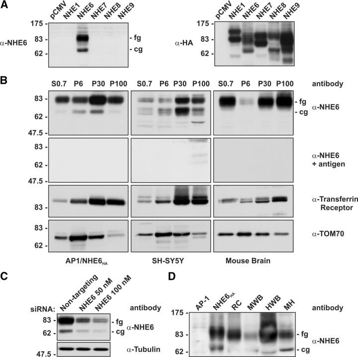
Characterization of an NHE6 isoform-specific rabbit polyclonal antibody by immunoblotting. A, Lysates of AP-1 cells transiently expressing rat NHE1HA, human NHE6HA, NHE7HA, NHE8HA, and NHE9HA were subjected to SDS-PAGE and immunoblot analysis. When the blot was probed with the affinity-purified anti-NHE6 antibody (α-NHE6), a signal was detected only in cells transfected with NHE6HA (left). The immunoblot was reprobed with a mouse monoclonal anti-HA antibody (α-HA), showing expression of all NHE isoforms (right). B, Homogenates of AP-1/NHE6HA cells, SH-SY5Y cells, and mouse whole-brain were fractionated by differential gravity centrifugation to enrich for the following subcellular compartments: S0.7 (total cell supernatant lacking nuclei), P6 (mitochondria), P30 (microsomes), and P100 (plasma membrane) (see Materials and Methods for details), followed by SDS-PAGE and immunoblot analysis with the anti-NHE6 antibody. NHE6 was enriched in the P30 microsomal and P100 plasma membrane fractions. When the NHE6 antibody was pre-incubated with the immunizing antigen, no bands were detected. The membranes were also immunoblotted with antibodies that recognize established compartment markers; i.e., the Tf-R, which is enriched in the endosomal P30 and the plasma membrane P100 fractions, and TOM70, which is enriched in the P6 mitochondrial fraction. C, In AP-1 cells stably expressing human NHE6HA, our NHE6 antibody detected reduced expression of NHE6 only in cells treated with the specific anti-NHE6 siRNA and not in cells treated with a scrambled siRNA (top). As a control, tubulin levels were not affected by the depletion of NHE6 by siRNA (bottom). D, Detection of native NHE6 in brain tissue homogenates. Lysates of AP-1 cells transiently transfected with empty vector (AP-1) or NHE6HA, as well as homogenates of rat cerebrum (RC), mouse whole-brain (MWB), human whole-brain (HWB), and mouse hippocampus (MH) were subjected to SDS-PAGE and immunoblot analysis with the anti-NHE6 antibody. The fully glycosylated (fg), mature form of NHE6, as well as the core-glycosylated form (cg) are visible.

