Figure 4.
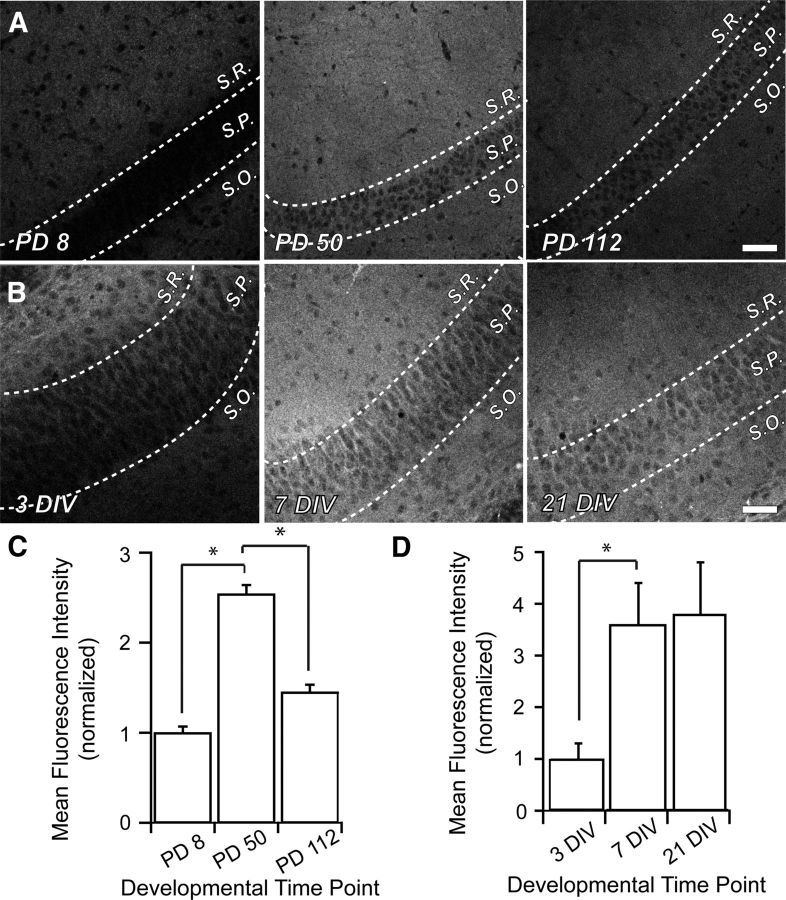
NHE6 is increased in area CA1 of the mouse hippocampus during synaptogenesis. Representative low-magnification confocal micrographs of NHE6 immunostaining in area CA1 of the mouse hippocampus. A, Cryostat-sectioned coronal brain slices from early postnatal (PD8; left), young adult (PD50; middle), and mature animals (PD112; right). During development, NHE6 puncta are diffuse throughout all strata of area CA1. Magnification in all parts (A, B) is identical. Scale bar, 50 μm. B, Organotypic mouse hippocampal slice cultures exhibited a similar expression profile as was detected in vivo. Throughout slice culture development (3 DIV, left; 7 DIV, middle; 21–28 DIV, right), NHE6 puncta are diffuse throughout all strata of area CA1. C, NHE6 significantly increases by 2.5-fold in area CA1 between PD8 and PD50 as measured by MFI. This period has been shown to be critical for elimination of superfluous connections in the brain and establishing refined synaptic circuits. There is also a significant decrease of NHE6 in area CA1 to ∼1.5× of early postnatal levels by PD112, by which time circuitry in the brain has stabilized. n = 5 slices for each time point; normalized MFI ± SEM. *p < 0.05 by Student's t test. D, NHE6, as measured by MFI, significantly increases in area CA1 during the period of synaptic pruning and refinement in organotypic hippocampal slice cultures, but does not return to early postnatal levels. n = 8 slices for each time point; normalized MFI ± SEM. *p < 0.05 by Student's t test. S.R., Stratum radiatum; S.P., stratum pyramidalis; S.O., stratum oriens; PD, postnatal day.

