Figure 6.

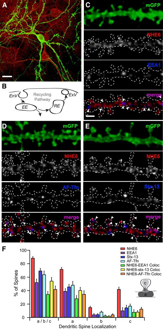
NHE6 colocalizes with early and recycling endosomal markers at dendritic spines of mouse primary hippocampal neurons. A, C–E, Representative confocal micrographs show immunofluorescent localization of NHE6 and its colocalization with early and recycling endosomes positive for EEA1, AF-Tfn, or Stx-13 in mouse primary hippocampal neurons (14+ DIV). Arrowheads denote protein localization at different regions of dendritic spines. A, NHE6 localizes to the somatodendritic compartment of an mGFP-positive neuron. It also localizes to neighboring unlabeled neurons and to cells of the surrounding glial cell bed. Scale bar, 20 μm. B, Simplified schematic representation of the endosomal recycling pathway at the dendritic spine surface. C, NHE6-EEA1 colocalization occurs at a subset of NHE6-positive dendritic spines in primary hippocampal neurons. EEA1 is almost exclusively found in close proximity to or partially overlapping NHE6 puncta. Magnification in all parts (C–E) is identical. Scale bar, 2 μm. D, mGFP-positive dendrite following incubation with AF-Tfn (100 μg/ml) for 1 h at 37°C to label recycling endosomes. Puncta of internalized AF-Tfn are frequently adjacent to or overlapping NHE6. E, NHE6-Stx-13 colocalization is seen only at a subset of NHE6-positive dendritic spines in primary hippocampal neurons. Stx-13 is almost exclusively found in close proximity or partially overlapping NHE6 puncta. F, Quantitative summary of NHE6 colocalization with recycling endosomal markers in dendritic spines of primary hippocampal neurons. NHE6 localizes to 90% of mature dendritic spines, showing colocalization with EEA1 at 35% of spines, AF-Tfn at 50% of spines, and with Stx-13 at 40% of spines. By the Mander's coefficient, the majority of Stx-13 and EEA1-positive puncta are significantly overlapped by NHE6 (Stx-13-NHE6: 0.8564, p < 0.05; EEA1-NHE6: 0.9611, p < 0.05), with approximately half of AF-Tfn overlapping with NHE6 (AF-Tfn-NHE6: 0.5733, p < 0.05). NHE6 colocalization with recycling endosomes occurs primarily at the spine base (a) and/or head (c), and to a minimal extent at the spine neck (b) (see inset schematic). Note: All immunopositive spines are described as a/b/c. Percentage values for dendritic spine areas (i.e., a, b, and c) do not add up to 100%, as individual spines could fall into more than one category. n = 163 dendritic spines from 172.78 μm dendrite from six neurons, nAF-Tfn = 221 dendritic spines from 228.70 μm dendrite from six neurons, 266 dendritic spines from 323.53 μm dendrite from eight neurons. Mean ± SEM localization per dendrite segment. White dashed line denotes secondary dendrite of an mGFP-positive dendrite. EnV, Endocytic vesicle; EE, early endosome; RE, recycling endosome; ExV, exocytic vesicle; EEA1, early endosome antigen 1; AF-Tfn, Alexa Fluor 568-conjugated transferrin; Stx-13, syntaxin-13; mGFP, membrane-tagged enhanced green fluorescent protein.
