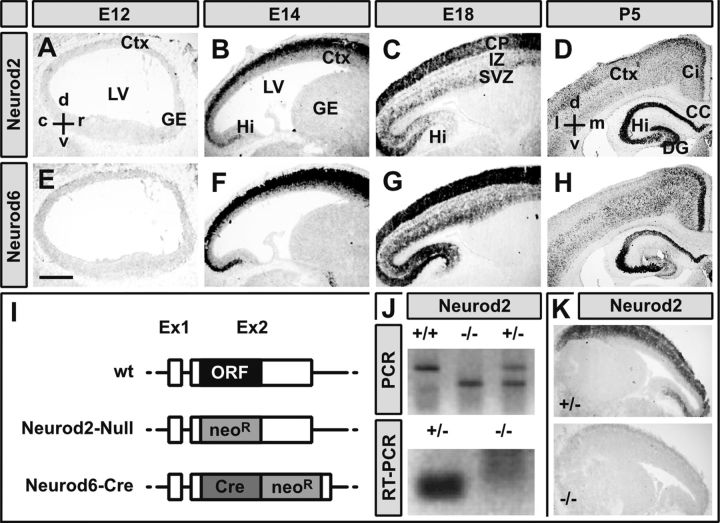
Figure 1.
Highly overlapping expression of Neurod2/6 during cortical development. A–H, Chromogenic in situ hybridization of immediately adjacent sagittal (A–C, E–G) and frontal (D, H) cryosections (14 μm) from mouse brains at indicated embryonic and postnatal stages with cRNA probes directed against Neurod2 (A–D) and Neurod6 (E–H) mRNA. I–K, Inactivation of the Neurod2 gene. Schematic drawings of a prototypic wild-type neuronal bHLH gene locus (wt) and constitutive mutant alleles of the Neurod2 (Neurod2–Null) and Neurod6 (Neurod6-Cre) gene (I). Inactivation of the Neurod2 gene was confirmed by PCR on genomic DNA (J), RT-PCR on total RNA from laser-dissected neocortex at P1 (J), and in situ hybridization at P1 (K). Scale bar, 500 μm. c, Caudal; d, dorsal; l, lateral; m, medial; r, rostral; v, ventral; Ci, cingulate cortex; Ctx, cerebral cortex; DG, dentate gyrus; GE, ganglionic eminence; Hi, hippocampus; IZ, intermediate zone; LV, lateral ventricle; neoR, neomycin resistance cassette.

