Figure 6.
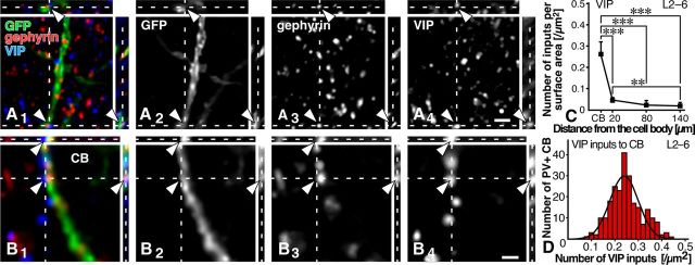
VIP inputs to PV neurons. A1–B4, GFP, gephyrin, and VIP were labeled with AF488 (green), AF568 (red), and AF647 (blue), respectively. VIP-positive axon terminals were frequently observed in close apposition to gephyrin immunoreactivity on the GFP-positive cell bodies, but less on the dendrites (arrowheads). C, The data obtained from all 32 dendrites and cell bodies were pooled. For the data in each layer, see Table 6. Each symbol represents mean ± SD. Statistical significance was judged by Tukey's multiple-comparison test after one-way ANOVA: **p < 0.01; ***p < 0.001. D, We randomly selected 20 PV neurons in each cortical layer of the S1 in three transgenic mice, and analyzed VIP inputs to the cell bodies of PV neurons (240 neurons in total). The frequency histogram against the input density was well fitted with a single Gaussian distribution (r = 0.94), compared with the mixture of Gaussian distributions, on the basis of the Bayesian information criterion. Scale bars: (in A4) A1–A3, 2 μm; (in B4) B1–B3, 1 μm.

