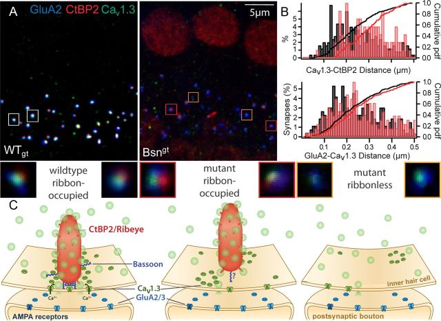
Figure 1.
Bassoon contributes to anchoring the ribbon at the presynaptic membrane. A, Rows of IHCs from a WTgt (left) and a Bsngt mutant (right) mouse immunostained with antibodies against CtBP2 (red, labeling synaptic ribbons and nuclei), Cav1.3 (green, labeling presynaptic calcium channels), and GluA2 (blue, labeling postsynaptic glutamate receptors, illustrating a normal number of synapses but reduced ribbon occupancy in Bsngt IHCs. B, Distances between the centers of mass of CtBP2- and Cav1.3-immunoreactive clusters (top) at Bsngt IHC synapses (red) were never as short as the closest ones in WTgt (black), suggesting disturbed ribbon anchorage to the presynaptic membrane. In contrast, the distance between Cav1.3 and GluA2-positive clusters (bottom) was comparable (from one representative pair of preparations). C, Schematic illustrations of individual ribbon synapses (sizes and numbers not to scale). In the normal IHC synapse (left), the AZ is occupied by a synaptic ribbon (red) tethered to the presynaptic membrane by Bassoon (dark blue) accompanied by ribbon-associated and cytosolic synaptic vesicles (light green). Presynaptic Ca2+ channels (dark green) are clustered in a row underneath the ribbon (Frank et al., 2010), and glutamate receptor clusters (blue) on the postsynaptic SGN boutons assume a ring-like shape (Meyer et al., 2009; Fig. 3D). Bassoon has an unknown tertiary structure with a central RIBEYE-binding site (tom Dieck et al., 2005) and two double zinc finger domains in the n-terminal region that target synaptic membranes (Dresbach et al., 2003). In Bsn mutant synapses, the ribbon can be present (middle) or absent (right), Ca2+ channels are reduced in number, and the clusters are disorganized, whereas glutamate receptor rings and SGN boutons appear to be mostly preserved. The separation between remaining Bsngt ribbons and the presynaptic membrane is increased. Insets, deconvolved higher-magnification images of representative synapses from A.

