Figure 3.
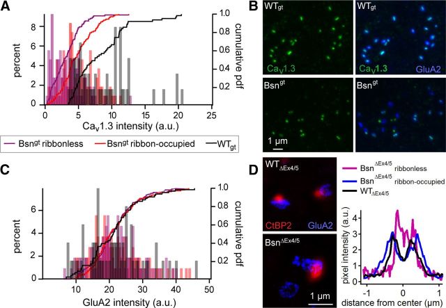
Altered presynaptic Ca2+ channel clusters and near normal postsynaptic AMPA receptor clusters at IHC synapses of Bsn mutants. A, Cav1.3 immunofluorescence in Bsngt IHCs (violet, n = 108 Bsngt ribbonless synapses; red, n = 108 Bsngt ribbon-occupied synapses) had reduced intensity compared with WTgt (black, n = 105 synapses). The analysis shown is derived from one representative pair of triple-stained samples as shown in Figure 1. Ribbon-occupied BSNgt synapses harbor more Cav1.3 immunofluorescence than ribbonless synapses but less than WTgt. B, Pair of double-stained preparations demonstrating the reduction in synaptic Cav1.3 (green), but not GluA2 receptor (blue), immunofluorescence. C, Distributions of GluA2 cluster immunofluorescence intensities of Bsngt and WTgt synapses overlapped (same preparation and color-codes as in A. D, Stimulated emission depletion microscopy demonstrating that ribbon-occupied and ribbonless synapses retained the ring-like morphology of GluA2/3 clusters (blue) in BsnΔEx4/5 synapses, albeit with a slightly smaller diameter in the absence of a presynaptic ribbon (CtBP2, red, confocal mode).

