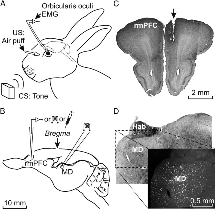
Figure 1.
Experimental design. A, For the classical conditioning of eyelid responses, animals were chronically implanted with EMG recording electrodes in the left orbicularis oculi muscle. The US consisted of an air puff presented to the ipsilateral cornea and the CS consisted of tones presented biaurally. B, Diagram of the rabbit brain illustrating the stimulating, recording, and injection sites. C, Photomicrograph illustrating the location of a stimulated site in the right rmPFC (arrow). D, Photomicrograph illustrating labeled neurons located in the MD nucleus after BDA injection in the rmPFC, as indicated in C. Calibrations for B–D are indicated. Hab, Habenula nucleus; MD, mediodorsal thalamic nucleus.

