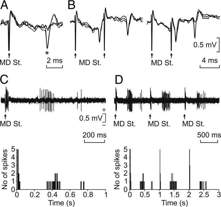
Figure 2.
Neuronal identification procedures. A, All-or-none antidromic activation of a rmPFC neuron by the stimulation of the ipsilateral mediodorsal thalamic nucleus (MD St.). B, Collision test for an rmPFC neuron activated from the MD nucleus. C, D, Synaptic activation of an identified rmPFC neuron represented with two different time bases. The stimulus consisted in a pair of pulses (100 μs in duration, 1 ms of interpulse interval, and up to 2 mA) presented at a rate of 1 Hz. The cumulative records for the synaptic activation of this neuron are represented in the two bottom graphs.

