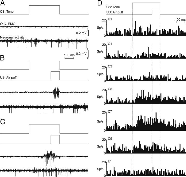
Figure 3.
Firing rate of rmPFC neurons during classical conditioning of eyelid responses. A, Representative examples of neuronal activity of rmPFC neurons and the electromyographic activity of the orbicularis oculi muscle (O.O. EMG). From top to bottom are illustrated CS presentation, the O.O. EMG, and the activity of an rmPFC neuron. Records were collected during the first habituation session. B, C, Similar sets of records (including air puff, US, presentation) collected during the second (B) and the fifth (C) conditioning sessions. Calibrations for A–C are indicated. D, Average of the firing rates (in spikes/s; sp/s) of rmPFC neurons recorded during the habituation (H1), conditioning (C1, C3, C5, C7, C9), and extinction (E1) sessions. The delay conditioning paradigm is indicated at the top. The dotted lines indicate the start and end of CS and US presentations. Note that a noticeable increase of neural firing in relation to the paired CS-US presentation took place only around the fifth-seventh sessions.

