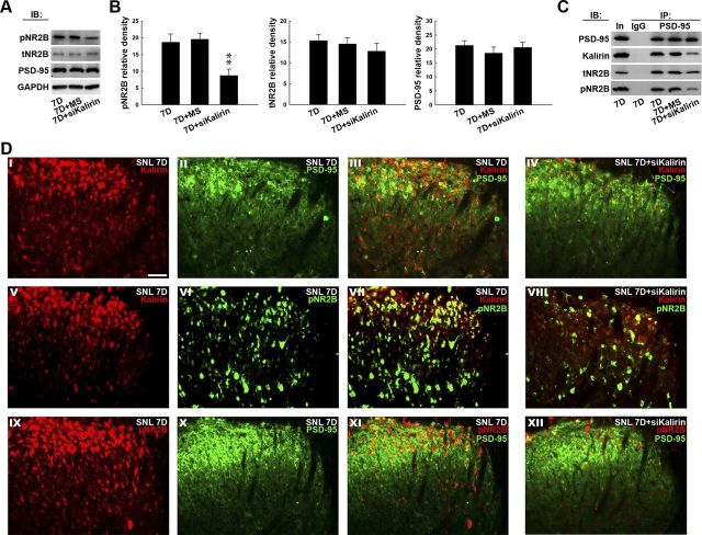
Figure 4.
Kalirin mRNA-targeting siRNA prevents NR2B phosphorylation and coupling with PSD-95. A, Representative Western blot showing the expression of pNR2B, tNR2B, and PSD-95 in response to the administration of siRNA that targeted kalirin mRNA. The lysates obtained from the ipsilateral dorsal horn 7 d after spinal nerve ligation were probed with antibodies against pNR2B, tNR2B, and PSD-95. The level of GAPDH was used as a loading control. IB, immunoblotting. B, Statistical analysis revealed that the spinal administration of kalirin-targeting siRNA (10 μg, 10 μl; 7D+siKalirin), but not missense siRNA (10 μg, 10 μl; 7D+MS), decreased the pNR2B band intensity compared with animals that received nerve ligation only (7D; **p < 0.01 vs 7D, n = 7). In contrast, neither treatment affected the tNR2B or PSD-95 band intensities (p > 0.05 vs 7D, n = 7). The data show mean ± SEM. C, Coimmunoprecipitation of PSD-95, kalirin, tNR2B, and pNR2B in the ipsilateral spinal cord. Dorsal horn samples obtained 7 d after operation were immunoprecipitated with antibodies against PSD-95 (IP: PSD-95) or control IgG (IP: IgG) and were then probed with PSD-95, kalirin, tNR2B, and pNR2B antibodies. In, input sample. The immunoreactivity probed by the kalirin-selective, tNR2B-selective, and pNR2B-selective antibodies in the PSD-95 immunoprecipitates was reduced by the kalirin mRNA-targeting siRNA, but not by the missense siRNA. No immunoreactivity was detected by PSD-95-selective, kalirin-selective, tNR2B-selective, and pNR2B-selective antibodies in the control IgG-recognized precipitates. In, input protein. D, Overlay images show the colocalized immunoreactivities of kalirin (I, red) with PSD-95 (II, green), kalirin (V, red), and pNR2B (VI, green), as well as pNR2B (IX, red) with PSD-95 (X, green) in the dorsal horn of the spinal slices obtained 7 d after spinal nerve ligation (SNL 7D; III, VII, and XI, yellow), which were remarkably reduced by kalirin mRNA-targeting siRNA (SNL 7D+siKalirin; IV, VIII, and XII, respectively). Each of these immunofluorescence images was replicated in seven sample preparations with similar results each time. Scale bar, 50 μm; thickness, 50 μm.

