
Figure 3.
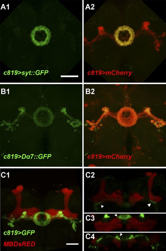
Distribution of presynaptic and postsynaptic regions in c819-EB neurons. A1, A2, Presynaptic regions labeled by Syt::GFP (green) mainly localized to the ring area of c819-EB neurons shown by mCherry (red) in (A2). B1, B2, Postsynaptic regions labeled by Dα7::GFP (green) distributed to the whole neuron shown by mCherry (red) in B2. C1–C4, MB247-DsRED (red) and c819>mGFP (green) showed MB and EB structures, respectively, which were close together in some regions (arrowheads). C1, Confocal projection of the brain frontal view. C2–C4, Single section of the volume view (C2 was a coronal section and C3, C4 were horizontal sections). Scale bars, 50 μm.