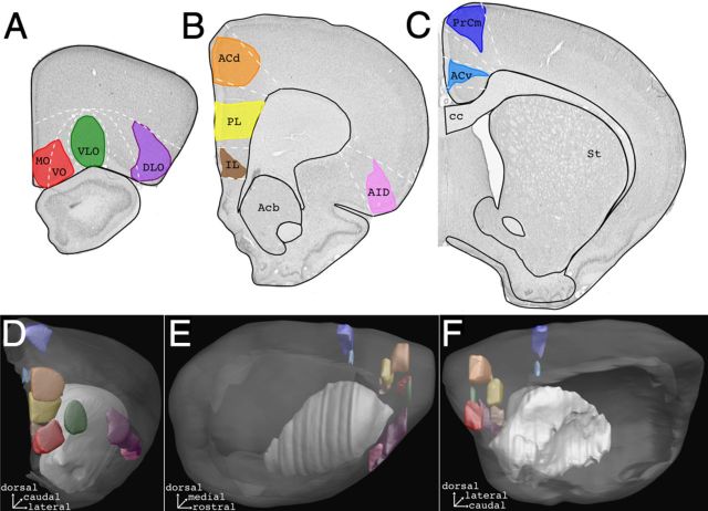
Figure 1.
Summary diagram of the anterograde tracer injection sites in the nine cases selected for 3D reconstruction of prefrontostriatal projections. A–C, Photomicrographs of coronal Nissl sections with delineation of the maximal extent of tracer. D–F, Position and extent of injection sites in the 3D model examined from a rostral view (D), lateral view (E), and medial view (F). Light gray, striatum; dark gray, cerebral cortex. ac, Anterior commissure; Acb, accumbens nucleus; cc, corpus callosum; St, striatum. Color codes: DLO (purple), dorsolateral orbital cortex; VLO (green), ventrolateral orbital cortex; PL (yellow), prelimbic cortex; MOVO (red), medial orbital and ventral orbital cortices; ACd (orange), dorsal anterior cingulate cortex; IL (brown), infralimbic cortex; ACv (cyan), ventral anterior cingulate cortex; AID (magenta), dorsal agranular insular cortex; PrCm (blue), medial precentral cortex.

