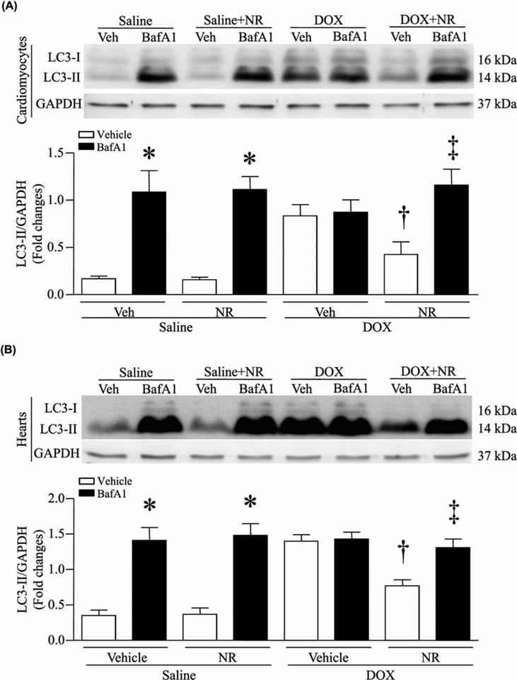
Figure 3. NR attenuated DOX-induced impairment of autophagic flux in cardiomyocytes.
(A) Cultured neonatal mouse cardiomyocytes were incubated with NR (500 μmol/l) or vehicle for 24 h, followed by DOX (DOX, 1 μmol/l) or saline in culture media for another 22 h. After that, bafilomycin A1 (BafA1) or vehicle (Veh) was added. About 2 h after addition of BafA1, western blot was performed to analyze the protein levels of LC3II and GAPDH. Upper panel: representative western blot for LC3II and GAPDH protein and lower panel: quantitation of LC3II/GAPDH ratio. (B) Mice received NR and 30 min later were injected with DOX or saline. About 5 days later after DOX injection, mice received BafA1 or Veh. About 2 h later, western blot was performed to analyze the protein levels of LC3II and GAPDH. Upper panel: representative western blot for LC3II and GAPDH protein and lower panel: quantitation of LC3II/GAPDH ratio. Data are mean + S.D. from four different cell cultures with each in duplication or five different hearts in each group. *P<0.05 versus saline + vehicle, †P<0.05 versus DOX + vehicle and ‡P<0.05 versus DOX + NR.

