
Figure 2.
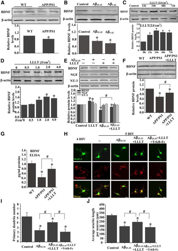
LLLT upregulates BDNF expression in cultured neurons derived from APP/PS1 transgenic mice and in Aβ-treated hippocampal neurons. A, Western blot analysis of hippocampal lysates from APP/PS1 transgenic mice and age-matched WT littermate mice at 6 months of age. B, Western blot analysis of BDNF expression in Aβ25–35-and Aβ1–42-treated primary hippocampal neurons. C, Representative Western blot assay for detecting the time-dependent effect of 2 J/cm2 LLLT on BDNF expression in SH-SY5Y cells. D, Representative Western blot assay for detecting the dose-dependent effect of LLLT on BDNF expression after 24 h. E, Western blot was performed to detect the BDNF, nerve growth factor (NGF), and NT-3 expression after Aβ25–35 treatment with or without LLLT in primary hippocampal neurons. F–G, BDNF expression was detected by Western blot (F) and ELISA (G) in hippocampal neurons derived from WT mice embryos or APP/PS1 embryos at 14 DIV with or without LLLT. H, Representative immunofluorescent images of 5 DIV hippocampal neurons under indicated treatments with MAP2 antibody to visualize dendrite (green). Staining with Rhodamine-labeled phalloidin to visualize F-actin (red). Scale bar, 10 μm. I, Effects of these treatments on the number of primary dendrites per neurons. For each group, >25 neurons were measured. J, Effects of these treatments on average dendritic length per neuron. For each group, >25 neurons were measured. All the data in these figures are presented as mean ± SEM four individual experiments. *p < 0.05 versus control group; #p < 0.05 versus indicated group.