
Figure 1.
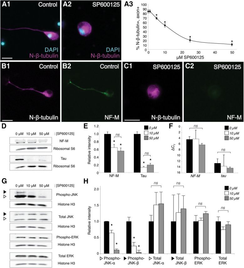
JNK inhibition compromises axon outgrowth and cytoskeletal protein expression. A1, A2, Representative images of neurons in control or SP600125-treated (50 μm) dissociated embryonic neural tube–myotome cultures, as indicated. Immunostaining for N-β-tubulin (magenta) labeled cell bodies and axons (if present), and DAPI (blue) labeled nuclei. A3, Quantification (30 neurons/culture, n = 6 cultures) of the percentage (±SEM) of N-β-tubulin+ cells that had axons in increasing concentrations of SP600125 indicated that JNK activity was required for axon initiation, reaching statistical significance at 5 μm SP600125 (p < 0.01, two-sided t-tests with control, 30 neurons/culture, n = 5 cultures). B, C, Double-label immunocytochemistry for N-β-tubulin (B1, C1, magenta) and NF-M (B2, C2, green), with or without 50 μm SP600125 (C1,C2 and B1,B2, respectively), showed a decrease in the level of NF-M expression when JNK was inhibited. Scale bars, 20 μm. D, Representative Western blots of cultures grown in 0, 10, or 50 μm SP600125. E, Intensities of NF-M and tau protein bands, after normalization against ribosomal protein S6 (loading control), were plotted relative to the intensity at 0 μm SP600125 (±SEM). JNK inhibition (10 and 50 μm) significantly reduced NF-M and tau levels compared with the 0 μm control (ns, nonsignificant; *p < 0.05 and p < 0.01 for NF-M and tau, respectively, one-way ANOVA with Tukey's post hoc test, n = 3 replicates of 25 pooled cultures for each condition); there was no significant difference in this effect between 10 and 50 μm SP600125 (p = 0.6, one-way ANOVA). F, qRT-PCR for NF-M and tau (ΔCT ± SD, relative to GAPDH) of cultures grown in 0, 10, or 50 μm SP600125 displayed no significant change in NF-M or tau RNA expression when JNK was inhibited (p = 0.5, one-way ANOVA, n = 3 replicates of 25 pooled cultures for each condition). G, H, Western blots of SP600125-treated cultures were probed with antibodies to activated (phospho-) JNK, total JNK, activated (phospho-) ERK, and total ERK to investigate effects of SP600125 on the levels of activated and total JNK versus activated and total ERK. Intensities of activated and total JNK and ERK bands, after normalization against histone H3, were plotted relative to the intensity at 0 μm SP600125 (±SEM). White and black triangles indicate the p46 (α) and p54 (β) isoforms of JNK, which in X. laevis are 44 and 48 kDa, respectively. Levels of activated JNK-α and JNK-β were significantly reduced (p < 0.01, one-way ANOVA with Tukey's post hoc test, n = 3 replicates of 25 pooled cultures for each condition) with SP600125 treatment, whereas no significant effects were observed for total JNK-α/β, activated ERK, and total ERK (p = 0.7, p = 0.9, p = 0.4, p = 0.3, respectively, one-way ANOVA), indicating that SP600125 specifically inhibited JNK activation.